Latest News

  • OCT 2023

    Qiancheng Awarded the 2023 Foresight Distinguished Student Award

    We are proud to announce that Qiancheng has received the 'Distinguished Student Award' from the Foresight Institute's Feynman Prizes, an esteemed recognition of his contributions to nanotechnology. His dedication and innovative work have significantly advanced our understanding in this field, showcasing the high calibre of research conducted in our lab. Congratulations, Qiancheng, on this outstanding achievement!

    image
  • AUG 2023

    Qiancheng Awarded the A*STAR International Fellowship

    We are thrilled to announce that Qiancheng has been awarded the prestigious A*STAR International Fellowship for post-doctoral training at the Wyss Institute, Harvard University – Congratulations Qiancheng! While we will greatly miss his contributions and presence in our lab, we are excited for the incredible journey and opportunities that await him in this next chapter of his career.

    image
  • AUG 2023

    Qiancheng Successfully Defended His Thesis

    We are proud to announce that Qiancheng has brilliantly defended his thesis titled ‘Functionalized DNA Nanostructures for Membrane Engineering,’ showcasing a remarkable depth of knowledge and innovation in his field. Congratulations on this outstanding achievement, Qiancheng! Your dedication and hard work have truly set a high standard in our lab.

    image
  • APR 2023

    Qi Awarded the 2023 Robert Dirks Molecular Programming Prize

    Dr. Qi Shen has been awarded the prestigious 2023 Robert Dirks Molecular Programming Prize for his groundbreaking work on constructing nuclear pore mimics using DNA origami. Established in honor of the late Dr. Robert Dirks, the prize recognizes outstanding achievements in the design, modeling, and implementation of complex molecular systems. Congratulations to Qi!

    image
  • APR 2023

    Minhwan Won the Best Poster Award at FNANO23

    Dr. Minhwan Chung presented his work with Dr. Kun Zhou at the 20th Annual Conference on the Foundations of Nanoscience (FNANO23) and won the Best Poster Award. Congratulations!

    image
  • APR 2023

    Eason Awarded the Prize Teaching Fellowship

    Eason has been awarded a Prize Teaching Fellowship for his outstanding teaching in 2022-2023. This award is one of the highest honors that a graduate student can attain at Yale. Congratulations!

    image
  • FEB 2023

    Congratulations!

    Qi successfully passed her qualifying exam!

  • DEC 2022

    Congratulations!

    Eason successfully passed his qualifying exam!

Past Events

  • APR 2022

    Congratulations!

    Longfei won an “Excellent contributed talk by a student or postdoc” award at the FNANO 2022 conference.

  • MAR 2022

    New Graduate Students

    Qi and Eason are starting as new graduate students in our lab, while Louis from France is joining us for an exchange. Welcome!

  • OCT 2021

    New Postdoctoral Fellow

    Kun Zhou is joining as a postdoctoral fellow. He was previously working in the Yonggang Ke lab at Emory University. Welcome!

  • JAN 2021

    New Students

    Ryan and Hanwen have joined the lab as undergraduate researchers. Welcome!

  • DEC 2020

    Congratulations to Our Recent Graduates

    John and Nathan have succesfully defended their theses. Congratulations to our new Dr. Powell and Dr. Williams!

  • JUN 2020

    New Postdoctoral Fellow

    Qingzhou is joining the Lin lab as a postdoctoral fellow. She recently graduated from the Hancock lab in Penn State University. Welcome!

  • SEP 2019

    New Exchange Student

    Taoran from the West China School of Stomatology, Sichuan University is joining us for an exchange. Welcome!

  • AUG 2019

    Congratulations to Ben!

    Ben successfully defended his thesis on "Selective gene-editing using an engineered Cas9 system and nuclei acid nanotechnology". Congratulations!

  • JUN 2019

    New Postdoctoral Fellow

    Longfei is joining the Lin lab as a postdoctoral fellow. He recently graduated from the Chengde Mao lab at Purdue University.

  • JUN 2019

    Congratulations to Michael!

    Michael has graduated with a Ph.D. in Cell Biology. He is joining the Farren Isaacs lab as a postdoctoral fellow. Congratulations!

  • FEB 2019

    Congratulations to Michael!

    Michael successfully defended his thesis on ”Vesicle Tubulation with Self-Assembling DNA Nanosprings: Biomimetic Nanotechnology toward the Re-capitulation & Re-purposing of Sub-cellular Functions within an Artificial Framework”. Congratulations!

  • DEC 2018

    New Student: Chun Xie

    Chun is a student from the Huazhong University of Science and Technology (HUST). He will be joining us on an one-year exchange program.

  • SEP 2018

    Farewell to Yang

    Yang will be leaving us to start his own lab in the School of Medicine, Shanghai Jiao Tong University. All the best!

  • AUG 2018

    New Exchange Student: Yan Cui

    Yan is a student from Tsinghua University. She will be joining us on an one-year exchange program.

  • JUL 2018

    New Exchange Student: Qi Yan

    Qi is a student from Tsinghua University. She will be joining us on an exchange program for two months.

  • JUN 2018

    Farewell to Zhao Zhang

    Zhao will be leaving us to join the Chapman Lab at the Department of Neuroscience, University of Wisconsin.

  • MAY 2018

    Congratulations to Mark

    Our former undergraduate student, Mark Zhu, graduated as the Class of 2018. Congratulations and all the best!

Current

  • image
    image

    Qi Shen

    Postdoctoral Fellow

    Qi Shen is currently an associate research scientist in the Department of Molecular Biophysics and Biochemistry and the Cell Biology Department of Yale University. He obtained his Ph.D. in physical chemistry from Peking University in 2013. During the Ph.D., he focused on the rational design of proteins and protein–protein interactions. Currently, he is working in the field of bottom‐up DNA nanodevice design and application. He has a multidisciplinary background and his research interests include DNA nanotechnology, synthetic biology, and structural cell biology.

  • image
    image

    Longfei Liu

    Postdoctoral Fellow

    Longfei graduated from University of Science and Technology of China (USTC) with a Bachelor’s Degree in Chemistry and received his Ph.D. degree at Purdue University. He is now a postdoctoral fellow in the Lin lab and works on DNA-nanotechnology-based membrane science. Outside the lab, he enjoys playing the piano, as well as outdoor activities including basketball and table tennis.

  • image
    image

    Qingzhou Feng

    Postdoctoral Fellow

    Qingzhou Feng graduated from Nankai University with a Bachelor’s degree in Biological Sciences and received her Ph.D. degree at Penn State University. She enjoys running, badminton and swimming.

  • image
    image

    Kun Zhou

    Postdoctoral Fellow

    To be updated. Stay tuned!

  • image
    image

    Qiancheng Xiong

    Graduate Student

    Qiancheng graduated from the University of Cambridge with a major in Genetics and worked as a Research Officer at the Institute of Molecular and Cell Biology (A*STAR, Singapore). He is a Ph.D. candidate in the Lin lab and works on engineering membrane encapsulated DNA origami structures. Outside the laboratory, he also experiments with caffeine extraction and yeast fermentation.

  • image
    image

    Qi Yan

    Graduate Student

    Qi graduated from the School of Life Sciences, Tsinghua University as a student in the Tsinghua Xuetang Program. She is now a student in Molecular Medicine, Pharmacology & Physiology track at Yale University. Qi is interested in measuring and manipulating membrane tension. She likes writing novels, reading comics, and drawing in her spare time.

  • image
    image

    Eason Cao

    Graduate Student

    Eason graduated from Fudan University with a Bachelor’s Degree in Pharmaceutical Sciences. His interest here is to apply DNA nanotechnology to deal with fundamental biological phenomena intractable through traditional methods. In his leisure time, he plays, composes, conducts music, and is passionate about all the world's arts and sciences. So, let’s talk about science and play Schubert sometime!

  • image
    image

    Ryan Bose-Roy

    Undergraduate Student

    Ryan is a first-year undergraduate in Trumbull College, originally from New York. He is really interested in using DNA origami techniques to build nanostructures with applications in medicine. In his spare time, he likes to draw, write, play viola, and swim.

Alumni

Patrick Fisher

PhD in Cell Biology (2017)

+ Follow

Zhao Zhang

Postdoctoral Fellow

+ Follow

Yang Yang

Postdoctoral Fellow

+ Follow

Michael Grome

PhD in Cell Biology (2019)

+ Follow

Ben Akhuetie-Oni

PhD in Molecular, Cellular and Developmental Biology (2019)

+ Follow

Chun Xie

Exchange Student (2019-2020)

Yan Cui

Exchange Student (2018-2019)

Taoran Tian

Exchange Student (2019-2020)

+ Follow

Nathan Williams

PhD in Cell Biology (2020)

John Powell

Postdoctoral Fellow

PhD in Cell Biology (2020)

Louis Bunel

Exchange Student (2022)

Publications

For a full list of Prof Lin's publications, please click here.

Filter by type:

Sort by year:

Recent Advances in DNA Origami-Engineered Nanomaterials and Applications

Pengfei Zhan, Andreas Peil, Qiao Jiang, Dongfang Wang, Shikufa Mousavi, Qiancheng Xiong, Qi Shen, Yingxu Shang, Baoquan Ding, Chenxiang Lin, Yonggang Ke, Na Liu
ReviewChemical Reviews, Volume 123, Issue 7, 29 March 2023, Pages 3976–4050

Abstract

DNA nanotechnology is a unique field, where physics, chemistry, biology, mathematics, engineering, and materials science can elegantly converge. Since the original proposal of Nadrian Seeman, significant advances have been achieved in the past four decades. During this glory time, the DNA origami technique developed by Paul Rothemund further pushed the field forward with a vigorous momentum, fostering a plethora of concepts, models, methodologies, and applications that were not thought of before. This review focuses on the recent progress in DNA origami-engineered nanomaterials in the past five years, outlining the exciting achievements as well as the unexplored research avenues. We believe that the spirit and assets that Seeman left for scientists will continue to bring interdisciplinary innovations and useful applications to this field in the next decade.

The capsid lattice engages a bipartite NUP153 motif to mediate nuclear entry of HIV-1 cores

Qi Shen, Sushila Kumari, Chaoyi Xu, Sooin Jang, Jiong Shi, Ryan C. Burdick, Lev Levintov, Qiancheng Xiong, Chunxiang Wu, Swapnil C. Devarkar, Taoran Tian, Therese N. Tripler, Yingxia Hu, Shuai Yuan, Joshua Temple, Qingzhou Feng, C. Patrick Lusk, Christopher Aiken, Alan N. Engelman, Juan R. Perilla, Vinay K. Pathak, Chenxiang Lin, Yong Xiong
Original ResearchProceedings of the National Academy of Sciences, Volume 120, Issue 13, 21 March 2023, Article e2202815120

Abstract

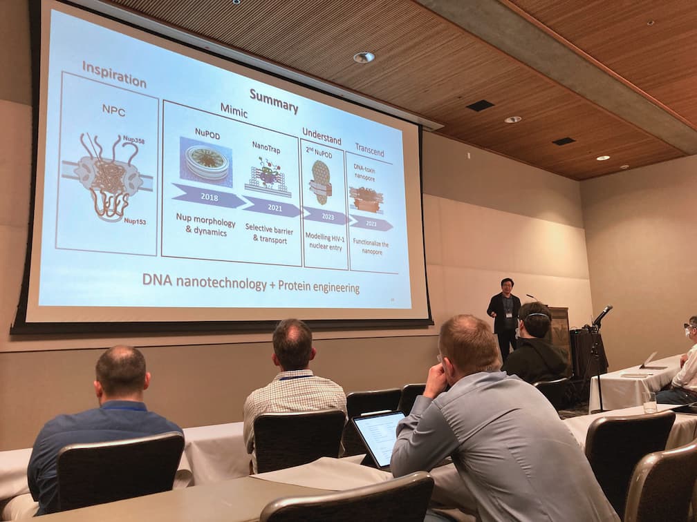
Increasing evidence has suggested that the HIV-1 capsid enters the nucleus in a largely assembled, intact form. However, not much is known about how the cone-shaped capsid interacts with the nucleoporins (NUPs) in the nuclear pore for crossing the nuclear pore complex. Here, we elucidate how NUP153 binds HIV-1 capsid by engaging the assembled capsid protein (CA) lattice. A bipartite motif containing both canonical and noncanonical interaction modules was identified at the C-terminal tail region of NUP153. The canonical cargo-targeting phenylalanine-glycine (FG) motif engaged the CA hexamer. By contrast, a previously unidentified triple-arginine (RRR) motif in NUP153 targeted HIV-1 capsid at the CA tri-hexamer interface in the capsid. HIV-1 infection studies indicated that both FG- and RRR-motifs were important for the nuclear import of HIV-1 cores. Moreover, the presence of NUP153 stabilized tubular CA assemblies in vitro. Our results provide molecular-level mechanistic evidence that NUP153 contributes to the entry of the intact capsid into the nucleus.

Modeling HIV-1 nuclear entry with nucleoporin-gated DNA-origami channels

Qi Shen, Qingzhou Feng, Chunxiang Wu, Qiancheng Xiong, Taoran Tian, Shuai Yuan, Jiong Shi, Gregory J. Bedwell, Ran Yang, Christopher Aiken, Alan N. Engelman, C. Patrick Lusk, Chenxiang Lin, Yong Xiong
Original ResearchNature Structural & Molecular Biology, 20 February 2023

Abstract

Delivering the virus genome into the host nucleus through the nuclear pore complex (NPC) is pivotal in human immunodeficiency virus 1 (HIV-1) infection. The mechanism of this process remains mysterious owing to the NPC complexity and the labyrinth of molecular interactions involved. Here we built a suite of NPC mimics—DNA-origami-corralled nucleoporins with programmable arrangements—to model HIV-1 nuclear entry. Using this system, we determined that multiple cytoplasm-facing Nup358 molecules provide avid binding for capsid docking to the NPC. The nucleoplasm-facing Nup153 preferentially attaches to high-curvature regions of the capsid, positioning it for tip-leading NPC insertion. Differential capsid binding strengths of Nup358 and Nup153 constitute an affinity gradient that drives capsid penetration. Nup62 in the NPC central channel forms a barrier that viruses must overcome during nuclear import. Our study thus provides a wealth of mechanistic insight and a transformative toolset for elucidating how viruses like HIV-1 enter the nucleus.

Functionalized DNA-Origami-Protein Nanopores Generate Large Transmembrane Channels with Programmable Size-Selectivity

Qi Shen, Qiancheng Xiong, Kaifeng Zhou, Qingzhou Feng, Longfei Liu, Taoran Tian, Chunxiang Wu, Yong Xiong, Thomas J. Melia, C. Patrick Lusk, Chenxiang Lin
Original ResearchJournal of the American Chemical Society, Volume 145, Issue 2, 18 January 2023, Pages 1292–1300

Abstract

The DNA-origami technique has enabled the engineering of transmembrane nanopores with programmable size and functionality, showing promise in building biosensors and synthetic cells. However, it remains challenging to build large (>10 nm), functionalizable nanopores that spontaneously perforate lipid membranes. Here, we take advantage of pneumolysin (PLY), a bacterial toxin that potently forms wide ring-like channels on cell membranes, to construct hybrid DNA–protein nanopores. This PLY-DNA-origami complex, in which a DNA-origami ring corrals up to 48 copies of PLY, targets the cholesterol-rich membranes of liposomes and red blood cells, readily forming uniformly sized pores with an average inner diameter of ∼22 nm. Such hybrid nanopores facilitate the exchange of macromolecules between perforated liposomes and their environment, with the exchange rate negatively correlating with the macromolecule size (diameters of gyration: 8–22 nm). Additionally, the DNA ring can be decorated with intrinsically disordered nucleoporins to further restrict the diffusion of traversing molecules, highlighting the programmability of the hybrid nanopores. PLY-DNA pores provide an enabling biophysical tool for studying the cross-membrane translocation of ultralarge molecules and open new opportunities for analytical chemistry, synthetic biology, and nanomedicine.

CLASP2 recognizes tubulins exposed at the microtubule plus-end in a nucleotide state–sensitive manner

Wangxi Luo, Vladimir Demidov, Qi Shen, Hugo Girão, Manas Chakraborty, Aleksandr Maiorov, Fazly I. Ataullakhanov, Chenxiang Lin, Helder Maiato, Ekaterina L. Grishchuk
Original ResearchScience Advances, Volume 9, Issue 1, 04 January 2023, eabq5404

Abstract

CLASPs (cytoplasmic linker-associated proteins) are ubiquitous stabilizers of microtubule dynamics, but their molecular targets at the microtubule plus-end are not understood. Using DNA origami–based reconstructions, we show that clusters of human CLASP2 form a load-bearing bond with terminal non-GTP tubulins at the stabilized microtubule tip. This activity relies on the unconventional TOG2 domain of CLASP2, which releases its high-affinity bond with non-GTP dimers upon their conversion into polymerization-competent GTP-tubulins. The ability of CLASP2 to recognize nucleotide-specific tubulin conformation and stabilize the catastrophe-promoting non-GTP tubulins intertwines with the previously underappreciated exchange between GDP and GTP at terminal tubulins. We propose that TOG2-dependent stabilization of sporadically occurring non-GTP tubulins represents a distinct molecular mechanism to suppress catastrophe at the freely assembling microtubule ends and to promote persistent tubulin assembly at the load-bearing tethered ends, such as at the kinetochores in dividing cells.

Actuating tension-loaded DNA clamps drives membrane tubulation

Longfei Liu, Qiancheng Xiong, Chun Xie, Frederic Pincet, Chenxiang Lin
Original ResearchScience Advances, Volume 8, Issue 41, 12 October 2022, eadd1830

Abstract

Membrane dynamics in living organisms can arise from proteins adhering to, assembling on, and exerting force on cell membranes. Programmable synthetic materials, such as self-assembled DNA nanostructures, offer the capability to drive membrane-remodeling events that resemble protein-mediated dynamics but with user-defined outcomes. An illustrative example is the tubular deformation of liposomes by DNA nanostructures with purposely designed shapes, surface modifications, and self-assembling properties. However, stimulus-responsive membrane tubulation mediated by DNA reconfiguration remains challenging. Here, we present the triggered formation of membrane tubes in response to specific DNA signals that actuate membrane-bound DNA clamps from an open state to various predefined closed states, releasing prestored energy to activate membrane deformation. We show that the timing and efficiency of vesicle tubulation, as well as the membrane tube widths, are modulated by the conformational change of DNA clamps, marking a solid step toward spatiotemporal control of membrane dynamics in an artificial system.

Omicron-specific mRNA vaccination alone and as a heterologous booster against SARS-CoV-2

Zhenhao Fang, Lei Peng, Renata Filler, Kazushi Suzuki, Andrew McNamara, Qianqian Lin, Paul A. Renauer, Luojia Yang, Bridget Menasche, Angie Sanchez, Ping Ren, Qiancheng Xiong, Madison Strine, Paul Clark, Chenxiang Lin, Albert I. Ko, Nathan D. Grubaugh, Craig B. Wilen, Sidi Chen
Original ResearchNature Communications, Volume 13, Issue 1, 06 June 2022, Page 3250

Abstract

The Omicron variant of SARS-CoV-2 recently swept the globe and showed high level of immune evasion. Here, we generate an Omicron-specific lipid nanoparticle (LNP) mRNA vaccine candidate, and test its activity in animals, both alone and as a heterologous booster to WT mRNA vaccine. Our Omicron-specific LNP-mRNA vaccine elicits strong antibody response in vaccination-naïve mice. Mice that received two-dose WT LNP-mRNA show a > 40-fold reduction in neutralization potency against Omicron than WT two weeks post boost, which further reduce to background level after 3 months. The WT or Omicron LNP-mRNA booster increases the waning antibody response of WT LNP-mRNA vaccinated mice against Omicron by 40 fold at two weeks post injection. Interestingly, the heterologous Omicron booster elicits neutralizing titers 10-20 fold higher than the homologous WT booster against Omicron variant, with comparable titers against Delta variant. All three types of vaccination, including Omicron alone, WT booster and Omicron booster, elicit broad binding antibody responses against SARS-CoV-2 WA-1, Beta, Delta variants and SARS-CoV. These data provide direct assessments of an Omicron-specific mRNA vaccination in vivo, both alone and as a heterologous booster to WT mRNA vaccine.

Frame-Guided Assembly of Amphiphiles

Yuanchen Dong, Yang Yang, Chenxiang Lin, Dongsheng Liu
ReviewAccounts of Chemical Research, Volume 55, Issue 14, 05 July 2022, Page 1938–1948

Abstract

Amphiphiles tend to self-assemble into various structures and morphologies in aqueous environments (e.g., micelles, tubes, fibers, vesicles, and lamellae). These assemblies and their properties have made significant impact in traditional chemical industries, e.g., increasing solubility, decreasing surface tension, facilitating foaming, etc. It is well-known that the molecular structure and its environment play a critical role in the assembly process, and many theories, including critical packing factor, thermodynamic models, etc., have been proposed to explain and predict the assembly morphology. It has been recognized that the morphology of the amphiphilic assembly plays important roles in determining the functions, such as curvature-dependent biophysical (e.g., liposome fusion and fission) and biochemical (e.g., lipid metabolism and membrane protein trafficking) processes, size-related EPR (enhanced permeability and retention) effects, etc. Meanwhile, various nanomaterials have promised great potential in directing the arrangement of molecules, thus generating unique functions. Therefore, control over the amphiphilic morphology is of great interest to scientists, especially in nanoscale with the assistance of functional nanomaterials. However, how to precisely manipulate the sizes and shapes of the assemblies is challenged by the entropic nature of the hydrophobic interaction. Inspired by the “cytoskeleton–membrane protein–lipid bilayer” principle of the cell membrane, a strategy termed “frame-guided assembly (FGA)” has been proposed and developed to direct the arrangement of amphiphiles. The FGA strategy welcomes various nanomaterials with precisely controlled properties to serve as scaffolds. By introducing scattered hydrophobic molecules, which are defined as either leading hydrophobic groups (LHGs) or nucleation seeds onto a selected scaffold, a discontinuous hydrophobic trace along the scaffold can be outlined, which will further guide the amphiphiles in the system to grow and form customized two- or three-dimensional (2D/3D) membrane geometries. Topologically, the supporting frame can be classified as three types including inner-frame, outer-frame, and planar-frame. Each type of FGA assembly possesses particular advantages: (1) The inner-frame, similar to endoskeletons of many cellular structures, steadily supports the membrane from the inside and exposes the full surface area outside. (2) The outer-frame, on the other hand, molds and constrains the membrane-wrapped vesicles to regulate their size and shape. It also allows postengineering of the frame to precisely decorate and dynamically manipulate the membrane. (3) The planar-frame mediates the growth of the 2D membrane that profits from the scanning-probe microscopic characterization and benefits the investigation of membrane proteins. In this Account, we introduce the recent progress of frame-guided assembly strategy in the preparation of customized amphiphile assemblies, evaluate their achievements and limitations, and discuss prospective developments and applications. The basic principle of FGA is discussed, and the morphology controllability is summarized in the inner-, outer-, and planar-frame categories. As a versatile strategy, FGA is able to guide different types of amphiphiles by designing specific LHGs for given molecular structures. The mechanism of FGA is then discussed systematically, including the driving force of the assembly, density and distribution of the LHGs, amphiphile concentration, and the kinetic process. Furthermore, the applications of FGA have been developed for liposome engineering, membrane protein incorporation, and drug delivery, which suggest the huge potential of FGA in fabricating novel and functional complexes.

Variant-specific vaccination induces systems immune responses and potent in vivo protection against SARS-CoV-2

Lei Peng, Paul A Renauer, Arya Ökten, Zhenhao Fang, Jonathan J Park, Xiaoyu Zhou, Qianqian Lin, Matthew B Dong, Renata Filler, Qiancheng Xiong, Paul Clark, Chenxiang Lin, Craig B Wilen, Sidi Chen
Original ResearchCell Reports Medicine, Volume 3, Issue 5, 17 May 2022, Page 100634

Abstract

Lipid nanoparticle (LNP)-mRNA vaccines offer protection against COVID-19; however, multiple variant lineages caused widespread breakthrough infections. Here, we generate LNP-mRNAs specifically encoding wild-type (WT), B.1.351, and B.1.617 SARS-CoV-2 spikes, and systematically study their immune responses. All three LNP-mRNAs induced potent antibody and T cell responses in animal models; however, differences in neutralization activity have been observed between variants. All three vaccines offer potent protection against in vivo challenges of authentic viruses of WA-1, Beta, and Delta variants. Single-cell transcriptomics of WT- and variant-specific LNP-mRNA-vaccinated animals reveal a systematic landscape of immune cell populations and global gene expression. Variant-specific vaccination induces a systemic increase of reactive CD8 T cells and altered gene expression programs in B and T lymphocytes. BCR-seq and TCR-seq unveil repertoire diversity and clonal expansions in vaccinated animals. These data provide assessment of efficacy and direct systems immune profiling of variant-specific LNP-mRNA vaccination in vivo.

Fluorogenic DNA-PAINT for faster, low-background super-resolution imaging

Kenny K. H. Chung, Zhao Zhang, Phylicia Kidd, Yongdeng Zhang, Nathan D. Williams, Bennett Rollins, Yang Yang, Chenxiang Lin, David Baddeley, Joerg Bewersdorf
Original ResearchNature Methods, Volume 19, 02 May 2022, Pages 554-559

Abstract

DNA-based points accumulation for imaging in nanoscale topography (DNA-PAINT) is a powerful super-resolution microscopy method that can acquire high-fidelity images at nanometer resolution. It suffers, however, from high background and slow imaging speed, both of which can be attributed to the presence of unbound fluorophores in solution. Here we present two-color fluorogenic DNA-PAINT, which uses improved imager probe and docking strand designs to solve these problems. These self-quenching single-stranded DNA probes are conjugated with a fluorophore and quencher at the terminals, which permits an increase in fluorescence by up to 57-fold upon binding and unquenching. In addition, the engineering of base pair mismatches between the fluorogenic imager probes and docking strands allowed us to achieve both high fluorogenicity and the fast binding kinetics required for fast imaging. We demonstrate a 26-fold increase in imaging speed over regular DNA-PAINT and show that our new implementation enables three-dimensional super-resolution DNA-PAINT imaging without optical sectioning.

DNA brick-assisted liposome sorting

Chenxiang Lin, Yang Yang
PatentUS Patent App. 17/162,008, 02 September 2021

Abstract

A method for producing uniform-size liposomes is provided. The liposomes are coated with a sorting agent to yield a plurality of density-modified liposomes of different sizes. These liposomes are then separated using a densitometric method. The sorting agent includes both a density-modifying moiety and a targeting moiety.

DNA-Origami NanoTrap for Studying the Selective Barriers Formed by Phenylalanine-Glycine-Rich Nucleoporins

Qi Shen, Taoran Tian, Qiancheng Xiong, Patrick D Ellis Fisher, Yong Xiong, Thomas J Melia, C Patrick Lusk, Chenxiang Lin
Original ResearchJournal of the American Chemical Society, Volume 143, Issue 31, 29 July 2021, Pages 12294-12303

Abstract

DNA nanotechnology provides a versatile and powerful tool to dissect the structure–function relationship of biomolecular machines like the nuclear pore complex (NPC), an enormous protein assembly that controls molecular traffic between the nucleus and cytoplasm. To understand how the intrinsically disordered, Phe-Gly-rich nucleoporins (FG-nups) within the NPC establish a selective barrier to macromolecules, we built a DNA-origami NanoTrap. The NanoTrap comprises precisely arranged FG-nups in an NPC-like channel, which sits on a baseplate that captures macromolecules that pass through the FG network. Using this biomimetic construct, we determined that the FG-motif type, grafting density, and spatial arrangement are critical determinants of an effective diffusion barrier. Further, we observed that diffusion barriers formed with cohesive FG interactions dominate in mixed-FG-nup scenarios. Finally, we demonstrated that the nuclear transport receptor, Ntf2, can selectively transport model cargo through NanoTraps composed of FxFG but not GLFG Nups. Our NanoTrap thus recapitulates the NPC’s fundamental biological activities, providing a valuable tool for studying nuclear transport.

FisB relies on homo-oligomerization and lipid binding to catalyze membrane fission in bacteria

Ane Landajuela, Martha Braun, Christopher D. A. Rodrigues, Alejandro Martínez-Calvo, Thierry Doan, Florian Horenkamp, Anna Andronicos, Vladimir Shteyn, Nathan D. Williams, Chenxiang Lin, Ned S. Wingreen, David Z. Rudner, Erdem Karatekin
Original ResearchPLoS Biology, Volume 19, Issue 6, 29 June 2021, e3001314

Abstract

Little is known about mechanisms of membrane fission in bacteria despite their requirement for cytokinesis. The only known dedicated membrane fission machinery in bacteria, fission protein B (FisB), is expressed during sporulation in Bacillus subtilis and is required to release the developing spore into the mother cell cytoplasm. Here, we characterized the requirements for FisB-mediated membrane fission. FisB forms mobile clusters of approximately 12 molecules that give way to an immobile cluster at the engulfment pole containing approximately 40 proteins at the time of membrane fission. Analysis of FisB mutants revealed that binding to acidic lipids and homo-oligomerization are both critical for targeting FisB to the engulfment pole and membrane fission. Experiments using artificial membranes and filamentous cells suggest that FisB does not have an intrinsic ability to sense or induce membrane curvature but can bridge membranes. Finally, modeling suggests that homo-oligomerization and trans-interactions with membranes are sufficient to explain FisB accumulation at the membrane neck that connects the engulfment membrane to the rest of the mother cell membrane during late stages of engulfment. Together, our results show that FisB is a robust and unusual membrane fission protein that relies on homo-oligomerization, lipid binding, and the unique membrane topology generated during engulfment for localization and membrane scission, but surprisingly, not on lipid microdomains, negative-curvature lipids, or curvature sensing.

Sorting sub-150-nm liposomes of distinct sizes by DNA-brick-assisted centrifugation

Yang Yang, Zhenyong Wu, Laurie Wang, Kaifeng Zhou, Kai Xia, Qiancheng Xiong, Longfei Liu, Zhao Zhang, Edwin R. Chapman, Yong Xiong, Thomas J. Melia, Erdem Karatekin, Hongzhou Gu, and Chenxiang Lin
Original ResearchNature Chemistry, Volume 13, Issue 4, 30 March 2021, Pages 335-342

Abstract

In cells, myriad membrane-interacting proteins generate and maintain curved membrane domains with radii of curvature around or below 50 nm. To understand how such highly curved membranes modulate specific protein functions, and vice versa, it is imperative to use small liposomes with precisely defined attributes as model membranes. Here, we report a versatile and scalable sorting technique that uses cholesterol-modified DNA ‘nanobricks’ to differentiate hetero-sized liposomes by their buoyant densities. This method separates milligrams of liposomes, regardless of their origins and chemical compositions, into six to eight homogeneous populations with mean diameters of 30–130 nm. We show that these uniform, leak-resistant liposomes serve as ideal substrates to study, with an unprecedented resolution, how membrane curvature influences peripheral (ATG3) and integral (SNARE) membrane protein activities. Compared with conventional methods, our sorting technique represents a streamlined process to achieve superior liposome size uniformity, which benefits research in membrane biology and the development of liposomal drug-delivery systems.

DNA Origami

Swarup Dey, Chunhai Fan, Kurt V. Gothelf, Jiang Li, Chenxiang Lin, Longfei Liu, Na Liu, Minke A. D. Nijenhuis, Barbara Saccà, Friedrich C. Simmel, Hao Yan, and Pengfei Zhan
ReviewNature Reviews Methods Primers, Volume 1, 2021, Article 13

Abstract

Biological materials are self-assembled with near-atomic precision in living cells, whereas synthetic 3D structures generally lack such precision and controllability. Recently, DNA nanotechnology, especially DNA origami technology, has been useful in the bottom-up fabrication of well-defined nanostructures ranging from tens of nanometres to sub-micrometres. In this Primer, we summarize the methodologies of DNA origami technology, including origami design, synthesis, functionalization and characterization. We highlight applications of origami structures in nanofabrication, nanophotonics and nanoelectronics, catalysis, computation, molecular machines, bioimaging, drug delivery and biophysics. We identify challenges for the field, including size limits, stability issues and the scale of production, and discuss their possible solutions. We further provide an outlook on next-generation DNA origami techniques that will allow in vivo synthesis and multiscale manufacturing.

DNA-Origami-Based Fluorescence Brightness Standards for Convenient and Fast Protein Counting in Live Cells

Nathan D. Williams, Ane Landajuela, Ravi Kiran Kasula, Wenjiao Zhou, John T. Powell, Zhiqun Xi, Farren J. Isaacs, Julien Berro, Derek Toomre, Erdem Karatekin, and Chenxiang Lin
Original ResearchNano Letters, Volume 20, Issue 12, 09 November 2020, Pages 8890-8896

Abstract

Fluorescence microscopy has been one of the most discovery-rich methods in biology. In the digital age, the discipline is becoming increasingly quantitative. Virtually all biological laboratories have access to fluorescence microscopes, but abilities to quantify biomolecule copy numbers are limited by the complexity and sophistication associated with current quantification methods. Here, we present DNA-origami-based fluorescence brightness standards for counting 5–300 copies of proteins in bacterial and mammalian cells, tagged with fluorescent proteins or membrane-permeable organic dyes. Compared to conventional quantification techniques, our brightness standards are robust, straightforward to use, and compatible with nearly all fluorescence imaging applications, thereby providing a practical and versatile tool to quantify biomolecules via fluorescence microscopy.

DNA Origami Post‐Processing by CRISPR‐Cas12a

Qiancheng Xiong, Chun Xie, Zhao Zhang, Longfei Liu, John T Powell, Qi Shen, Chenxiang Lin
Original ResearchAngewandte Chemie International Edition in English, Volume 59, Issue 10, 02 March 2020, Pages 3956-3960

Abstract

Customizable nanostructures built through the DNA‐origami technique hold tremendous promise in nanomaterial fabrication and biotechnology. Despite the cutting‐edge tools for DNA‐origami design and preparation, it remains challenging to separate structural components of an architecture built from—thus held together by—a continuous scaffold strand, which in turn limits the modularity and function of the DNA‐origami devices. To address this challenge, here we present an enzymatic method to clean up and reconfigure DNA‐origami structures. We target single‐stranded (ss) regions of DNA‐origami structures and remove them with CRISPR‐Cas12a, a hyper‐active ssDNA endonuclease without sequence specificity. We demonstrate the utility of this facile, selective post‐processing method on DNA structures with various geometrical and mechanical properties, realizing intricate structures and structural transformations that were previously difficult to engineer. Given the biocompatibility of Cas12a‐like enzymes, this versatile tool may be programmed in the future to operate functional nanodevices in cells.

RNA returns to the fold

Qi Shen, Chenxiang Lin
CommentaryNature Chemistry, Volume 12, Issue 3, 27 February 2020, Pages 221-222

Abstract

RNA has multiple roles in biology, enabled by its structural diversity. Now, artificially grafted RNA motifs have been encoded in a single RNA strand to form self-assembling nanostructures with controlled geometry and function.

Engineering Lipid Membranes with Programmable DNA Nanostructures

Qi Shen, Michael W Grome, Yang Yang, Chenxiang Lin
ReviewAdvanced Biosystems, Volume 4, Issue 1, 13 January 2020, Page 1900215

Abstract

Lipid and DNA are abundant biomolecules with critical functions in cells. The water‐insoluble, amphipathic lipid molecules are best known for their roles in energy storage (e.g., as triglyceride), signaling (e.g., as sphingolipid), and compartmentalization (e.g., by forming membrane‐enclosed bodies). The soluble, highly negatively charged DNA, which stores the cells' genetic information, has proven to be an excellent material for constructing programmable nanostructures in vitro thanks to its self‐assembling capabilities. These two seemingly distant molecules make contact within cell nuclei, often via lipidated proteins, with proposed functions of modulating chromatin structures. Carefully formulated lipid/DNA complexes are promising reagents for gene therapy. The past few years have seen an emerging research field of interfacing DNA nanostructures with lipid membranes, with an overarching goal of generating DNA/lipid hybrid materials that possess novel and controllable structure, dynamics, and function. An arsenal of DNA‐based tools has been created to coat, mold, deform, and penetrate lipid bilayers, affording the ability to manipulate membranes with nanoscopic precision. These membrane engineering methods not only enable quantitative biophysical studies, but also open new opportunities in synthetic biology (e.g., artificial cells) and therapeutics (e.g., drug delivery).

A Programmable DNA-Origami Platform for Studying Lipid Transfer between Bilayers

Xin Bian, Zhao Zhang, Qiancheng Xiong, Pietro De Camilli, Chenxiang Lin
Original ResearchNature Chemical Biology, Volume 15, Issue 8, 18 July 2019, Pages 830–837

Abstract

Non-vesicular lipid transport between bilayers at membrane contact sites plays important physiological roles. Mechanistic insight into the action of lipid-transport proteins localized at these sites requires determination of the distance between bilayers at which this transport can occur. Here we developed DNA-origami nanostructures to organize size-defined liposomes at precise distances and used them to study lipid transfer by the synaptotagmin-like mitochondrial lipid-binding protein (SMP) domain of extended synaptotagmin 1 (E-Syt1). Pairs of DNA-ring-templated donor and acceptor liposomes were docked through DNA pillars, which determined their distance. The SMP domain was anchored to donor liposomes via an unstructured linker, and lipid transfer was assessed via a Förster resonance energy transfer (FRET)-based assay. We show that lipid transfer can occur over distances that exceed the length of an SMP dimer, which is compatible with the shuttle model of lipid transport. The DNA nanostructures developed here can also be adapted to study other processes occurring where two membranes are closely apposed to each other.

Stiffness and Membrane Anchor Density Modulate DNA-Nanospring-Induced Vesicle Tubulation

Michael W Grome, Zhao Zhang, Chenxiang Lin
Original ResearchACS Applied Materials & Interfaces, Volume 11, Issue 26, 21 June 2019, Pages 22987-22992

Abstract

DNA nanotechnology provides an avenue for the construction of rationally designed artificial assemblages with well-defined and tunable architectures. Shaped to mimic natural membrane-deforming proteins and equipped with membrane anchoring molecules, curved DNA nanostructures can reproduce subcellular membrane remodeling events such as vesicle tubulation in vitro. To systematically analyze how structural stiffness and membrane affinity of DNA nanostructures affect the membrane remodeling outcome, here we build DNA-origami curls with varying thickness and amphipathic peptide density, and have them polymerize into nanosprings on the surface of liposomes. We find that modestly reducing rigidity and maximizing the number of membrane anchors not only promote membrane binding and remodeling but also lead to the formation of lipid tubules with better defined diameters, highlighting the ability of programmable DNA-based constructs to controllably deform the membrane.

Quantification of Biomolecular Dynamics Inside Real and Synthetic Nuclear Pore Complexes Using Time-Resolved Atomic Force Microscopy

George J. Stanley, Bernice Akpinar, Qi Shen, Patrick D. Ellis Fisher, C. Patrick Lusk, Chenxiang Lin, Bart W. Hoogenboom
Original ResearchACS Nano, Volume 13, Issue 7, 26 June 2019, Pages 7949-7956

Abstract

Over the past decades, atomic force microscopy (AFM) has emerged as an increasingly powerful tool to study the dynamics of biomolecules at nanometer length scales. However, the more stochastic the nature of such biomolecular dynamics, the harder it becomes to distinguish them from AFM measurement noise. Rapid, stochastic dynamics are inherent to biological systems comprising intrinsically disordered proteins. One role of such proteins is in the formation of the transport barrier of the nuclear pore complex (NPC): the selective gateway for macromolecular traffic entering or exiting the nucleus. Here, we use AFM to observe the dynamics of intrinsically disordered proteins from two systems: the transport barrier of native NPCs and the transport barrier of a mimetic NPC made using a DNA origami scaffold. Analyzing data recorded with 50-200 ms temporal resolution, we highlight the importance of drift correction and appropriate baseline measurements in such experiments. In addition, we describe an autocorrelation analysis to quantify time scales of observed dynamics and to assess their veracity-an analysis protocol that lends itself to the quantification of stochastic fluctuations in other biomolecular systems. The results reveal the surprisingly slow rate of stochastic, collective transitions inside mimetic NPCs, highlighting the importance of FG-nup cohesive interactions.

Vesicle Tubulation with Self‐Assembling DNA Nanosprings

Michael W. Grome, Zhao Zhang, Frédéric Pincet, Chenxiang Lin
Original ResearchAngewandte Chemie International Edition in English, Volume 57, Issue 19, 04 May 2018, Pages 5330-5334

Abstract

A major goal of nanotechnology and bioengineering is to build artificial nanomachines capable of generating specific membrane curvatures on demand. Inspired by natural membrane‐deforming proteins, we designed DNA‐origami curls that polymerize into nanosprings and show their efficacy in vesicle deformation. DNA‐coated membrane tubules emerge from spherical vesicles when DNA‐origami polymerization or high membrane‐surface coverage occurs. Unlike many previous methods, the DNA self‐assembly‐mediated membrane tubulation eliminates the need for detergents or top‐down manipulation. The DNA‐origami design and deformation conditions have substantial influence on the tubulation efficiency and tube morphology, underscoring the intricate interplay between lipid bilayers and vesicle‐deforming DNA structures.

A Programmable DNA Origami Platform for Organizing Intrinsically Disordered Nucleoporins within Nanopore Confinement

Patrick D. Ellis Fisher, Qi Shen, Bernice Akpinar, Luke K. Davis, Kenny Kwok Hin Chung, David Baddeley, Anđela Šarić, Thomas J. Melia, Bart W. Hoogenboom, Chenxiang Lin, C. Patrick Lusk
Original Research ACS Nano, Volume 12, Issue 2, 19 January 2018, Pages 1508-1518

Abstract

Nuclear pore complexes (NPCs) form gateways that control molecular exchange between the nucleus and the cytoplasm. They impose a diffusion barrier to macromolecules and enable the selective transport of nuclear transport receptors with bound cargo. The underlying mechanisms that establish these permeability properties remain to be fully elucidated but require unstructured nuclear pore proteins rich in Phe-Gly (FG)-repeat domains of different types, such as FxFG and GLFG. While physical modeling and in vitro approaches have provided a framework for explaining how the FG network contributes to the barrier and transport properties of the NPC, it remains unknown whether the number and/or the spatial positioning of different FG-domains along a cylindrical, ∼40 nm diameter transport channel contributes to their collective properties and function. To begin to answer these questions, we have used DNA origami to build a cylinder that mimics the dimensions of the central transport channel and can house a specified number of FG-domains at specific positions with easily tunable design parameters, such as grafting density and topology. We find the overall morphology of the FG-domain assemblies to be dependent on their chemical composition, determined by the type and density of FG-repeat, and on their architectural confinement provided by the DNA cylinder, largely consistent with here presented molecular dynamics simulations based on a coarse-grained polymer model. In addition, high-speed atomic force microscopy reveals local and reversible FG-domain condensation that transiently occludes the lumen of the DNA central channel mimics, suggestive of how the NPC might establish its permeability properties.

Directing reconfigurable DNA nanoarrays

Yang Yang, Chenxiang Lin
Commentary Science, Volume 357, Issue 6349, 28 July 2017, Pages 352-353

Abstract

The ability to faithfully pass information in a cascaded and controllable fashion has worked wonders for civilization and biology (see the figure). In the molecular engineering enterprise, researchers have craved a similar level of control over information flow within a network at nanometer-to-micrometer scale. On page 371 of this issue, Song et al. have used DNA, a readily available biopolymer with well-established structure and association rules, to construct finite-sized nanoarrays that transmit information from one side to the other in the form of structural transformation (1). This marks an important step toward programming nanoscale motions because it provides a generalizable way to propagate a local structural change across long distances along designated pathways.

Placing and shaping liposomes with reconfigurable DNA nanocages

Zhao Zhang, Yang Yang, Frederic Pincet, Marc C. Llaguno, Chenxiang Lin
Original Research Nature Chemistry, Volume 9, Issue 7, 23 June 2017, Pages 653-659

Abstract

The diverse structure and regulated deformation of lipid bilayer membranes are among a cell's most fascinating features. Artificial membrane-bound vesicles, known as liposomes, are versatile tools for modelling biological membranes and delivering foreign objects to cells. To fully mimic the complexity of cell membranes and optimize the efficiency of delivery vesicles, controlling liposome shape (both statically and dynamically) is of utmost importance. Here we report the assembly, arrangement and remodelling of liposomes with designer geometry: all of which are exquisitely controlled by a set of modular, reconfigurable DNA nanocages. Tubular and toroid shapes, among others, are transcribed from DNA cages to liposomes with high fidelity, giving rise to membrane curvatures present in cells yet previously difficult to construct in vitro. Moreover, the conformational changes of DNA cages drive membrane fusion and bending with predictable outcomes, opening up opportunities for the systematic study of membrane mechanics.

DNA Origami Rotaxanes: Tailored Synthesis and Controlled Structure Switching

John T. Powell, Benjamin O. Akhuetie‐Oni, Zhao Zhang, Chenxiang Lin
Original ResearchAngewandte Chemie International Edition in English, Volume 55, Issue 38, 12 September 2016, Pages 11412-11416

Abstract

Mechanically interlocked supramolecular assemblies are appealing building blocks for creating functional nanodevices. Herein, we describe the multistep assembly of large DNA origami rotaxanes that are capable of programmable structural switching. We validated the topology and structural integrity of these rotaxanes by analyzing the intermediate and final products of various assembly routes by electrophoresis and electron microscopy. We further analyzed two structure‐switching behaviors of our rotaxanes, which are both mediated by DNA hybridization. In the first mechanism, the translational motion of the macrocycle can be triggered or halted at either terminus. In the second mechanism, the macrocycle can be elongated after completion of the rotaxane assembly, giving rise to a unique structure that is otherwise difficult to access.

Self-assembly of size-controlled liposomes on DNA nanotemplates

Yang Yang, Jing Wang, Hideki Shigematsu, Weiming Xu, William M. Shih, James E. Rothman, Chenxiang Lin
Original ResearchNature Chemistry, Volume 8, Issue 5, 21 March 2016, Pages 476-483

Abstract

Artificial lipid-bilayer membranes are valuable tools for the study of membrane structure and dynamics. For applications such as the study of vesicular transport and drug delivery, there is a pressing need for artificial vesicles with controlled size. However, controlling vesicle size and shape with nanometre precision is challenging, and approaches to achieve this can be heavily affected by lipid composition. Here, we present a bio-inspired templating method to generate highly monodispersed sub-100-nm unilamellar vesicles, where liposome self-assembly was nucleated and confined inside rigid DNA nanotemplates. Using this method, we produce homogeneous liposomes with four distinct predefined sizes. We also show that the method can be used with a variety of lipid compositions and probe the mechanism of templated liposome formation by capturing key intermediates during membrane self-assembly. The DNA nanotemplating strategy represents a conceptually novel way to guide lipid bilayer formation and could be generalized to engineer complex membrane/protein structures with nanoscale precision.

Our Lab

You can find us at our lab located at the Nanobiology Institute on the Yale West Campus.

  850 West Campus Drive, ISTC #213C, West Haven, CT 06516

There is a free Yale University Shuttle service (Purple Line & Green Line) between the West Campus and the main campus in New Haven.

You may click here for more information.

Our PI

You may also contact Prof Lin directly via email or phone.

  •    office: 203-737-3924
  •    fax: 203-737-3212
  •    chenxiang.lin@yale.edu